All topics

Explain how infection by a pathogen can be detected by an ELISA test for antigens.

Explain how cells and cell components in the blood defend the body against infectious disease.

Outline reasons for the therapeutic use of stem cells.

If the heart of a vertebrate is removed and placed in a solution of nutrients, it will continue to beat for many hours. Related to this, which of the following is true?

Describe briefly the endosymbiotic theory.

Outline how photosynthesis produces glucose.

Discuss the control of blood glucose levels and the consequences if they are not maintained.

The graph below shows the levels of hormones during the menstrual cycle. 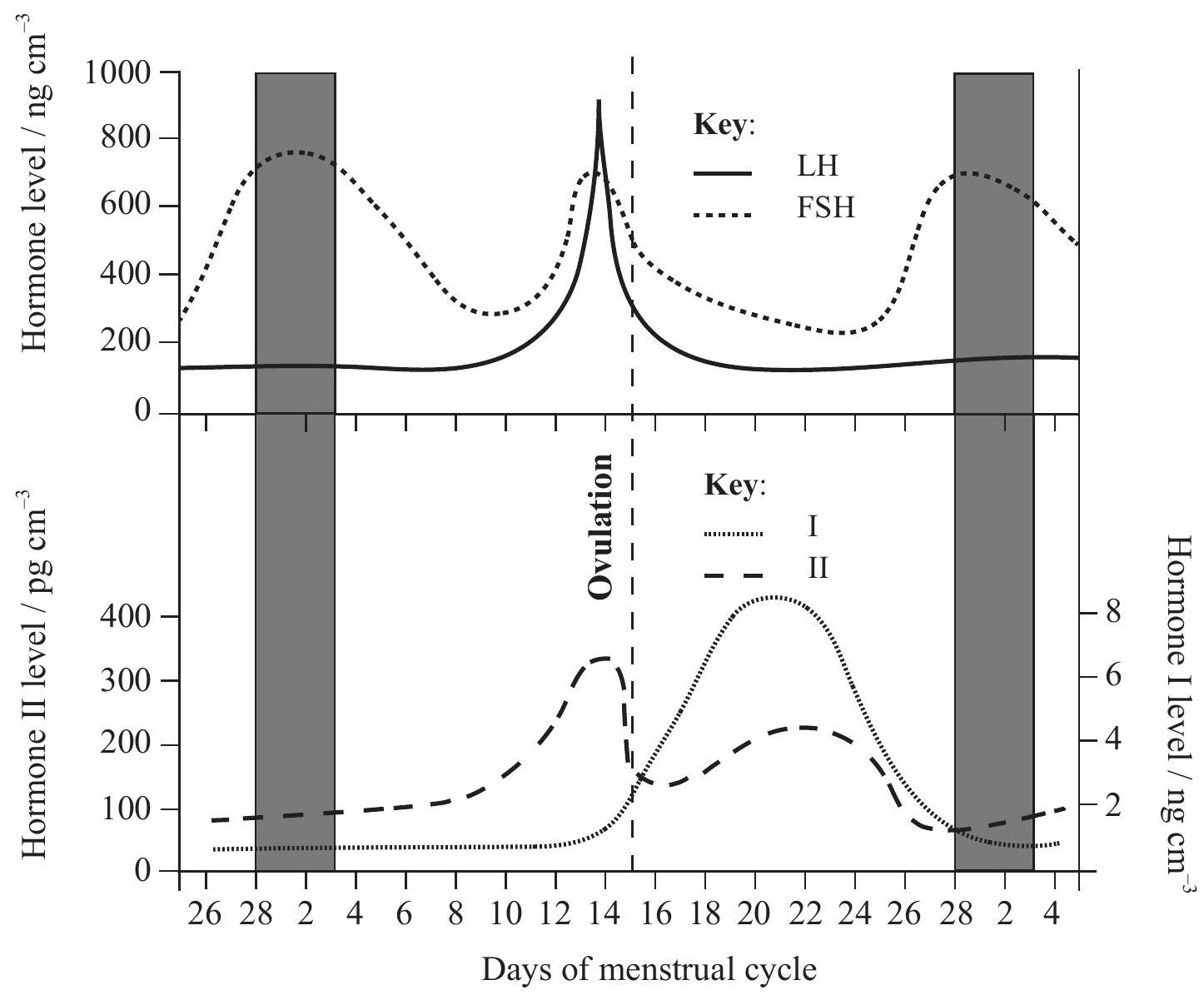

Identify hormones I and II.

Outline the roles of FSH in the menstrual cycle.

FSH is secreted by the pituitary gland. During pregnancy, FSH secretion is inhibited. Suggest how FSH secretion could be inhibited during pregnancy.

Draw a labelled diagram of the human heart showing the attached blood vessels.

Describe the action of the heart in pumping blood.

Nerves connecting the brain and heart contain neurons that control heart rate. Explain how a nerve message passes from one neuron to another neuron.

The graph shows the amounts of two substances present in food ingested by a healthy person as it moves along the gut.
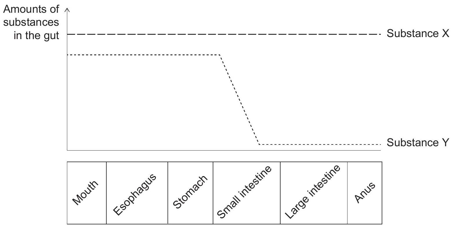
Which substances could X and Y be?

In 1628, the physician William Harvey described details of the circulation of blood for the first time. In one experiment, he tied a tight bandage around the upper arm of a volunteer to display the blood vessels in the lower arm more clearly. He pressed his finger on the blood vessel at H. At the same time, he pushed the blood in the vessel from H to O with a second finger, removing the blood as shown in the diagram. When the finger at H was released, the blood vessel refilled with blood.  [Source: adapted from William Harvey original plate]

Identify the type of blood vessels shown in the diagram.

Deduce what the experiment demonstrated about the circulation of blood.

What is the name and source of the hormone that regulates basal metabolic rate?
| Name | Source | |
|---|---|---|
| A. | ADH | kidneys |
| B. | melatonin | pineal gland |
| C. | thyroxin | thyroid gland |
| D. | glucagon | pancreas |

The structure of part of the digestive system is shown in the diagram below. 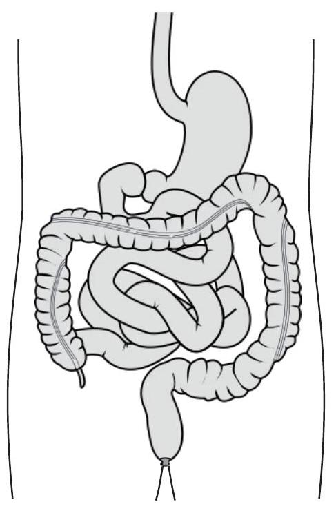 [Source: https://wccyusoueahdpdicsyrg.supabase.co/storage/v1/object/public/question-images/images/69c87bb4-31dd-4a17-a292-cd2edda2d48f.jpg Created by Wikipedia user: Madhero88.]

Label the diagram to show the structure that is involved in digestion of proteins in acid conditions (using the letter A).

Label the diagram to show the structure where most absorption of water to prevent dehydration occurs (using the letter B).

Label the diagram to show the structure where most absorption of nutrients occurs (using the letter C).

Explain how the structure of veins is adapted to their function.

Cells defend the body against pathogens. Outline how some of these cells ingest pathogens in the blood and in body tissues.
